

When my son started having seizures just 30 hours after he was born, an MRI a few days later showed us that he had hemimegalencephaly – a massive brain malformation that affected one side of his brain. It was huge, and there was no doubt in anyone’s mind that it was there.
Although I felt like my world was ending, looking back, I can’t help but think how lucky we were. We had answers right away and knew early on that the only treatment to attempt to stop his drug-resistant seizures was hemispherectomy, a type of resective epilepsy surgery where half the brain is removed.
Other parents of children with drug-resistant seizures aren’t as fortunate. After countless tests, the MRI comes back “normal” and the child is not a candidate for traditional, resective epilepsy surgery or responsive neurostimulation in the brain’s cerebral cortex. One hidden cause of seizures is a type of brain deformity called bottom-of-sulcus dysplasia. These tiny abnormalities hide deep in the folds of the brain, making difficult even for expert radiologists to see on standard MRI scans. Missing them matters because if they’re found, these lesions can often be surgically removed or destroyed with fine lasers, sometimes leading to seizure freedom.
I need to pause here to be clear: I am not a scientist or a medical doctor. I am a parent and patient advocate sharing what I have learned, and I hope I am summarizing the research correctly. If you have questions about your child’s care, please talk with your medical team. They are the best source of guidance for your family. And if you are a clinician or researcher and notice I’ve misunderstood something, please email me and let me know.
About the brain’s cortex
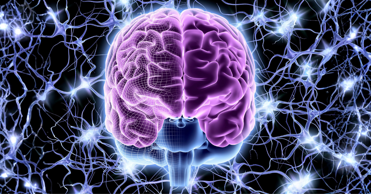
You’re probably most familiar with the images of the brain that show its folds and grooves. The foldy, groovy part is called the cortex and is the outer layer of the brain. The top of a fold is called the gyrus and the bottom the sulcus.

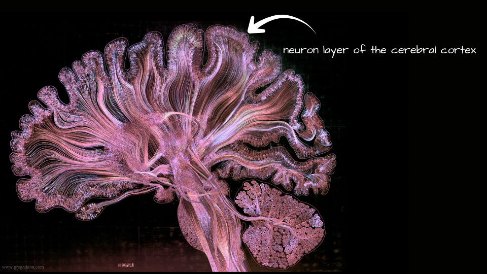
The grooves and folds of the outer layer of the brain are where many of the brain’s neurons are located. Artist and neuroscientist Dr. Greg Dunn does some spectacular microetchings using images from real brain MRIs, like this image that shows about 500,000 neurons. The fuzzy, mossy-looking part along the edges are the layers of neurons which as you can see are in the folds and grooves.
This simple drawing shows us these folds and grooves in general detail. The gray matter is where the neurons are located and where the cortical dysplasia can hide.

Now let’s get up close. You can see there are a lot of neurons in the folds and grooves – about 70,000 – 90,000 per square millimeter! It’s easy to see how it can be hard to find cortical dysplasia, which is basically deformed neurons or neurons that are not located in the right spot.

This is a basic structural MRI of the brain. Although you can see the folds and grooves, it’s hard to see detail.

When seizures resist medication, doctors may order an epilepsy-protocol MRI. This scan uses higher resolution settings to search for tiny abnormalities. Compared to a standard MRI, it uses thinner image slices and concentrates on imaging parts of the brain where seizures often start, such as the hippocampus and cortex.
Unfortunately, even epilepsy protocol MRI can miss subtle brain lesions. This video does a nice job of explaining it:
What is Bottom-of-Sulcus Dysplasia?
Bottom-of-sulcus dysplasia is a subtle malformation of brain tissue located in the bottom of a sulcus (a groove on the brain’s surface). Because of its size and location, it can be difficult to see even with epilepsy protocol MRI, leaving families in limbo. It is a known cause of focal epilepsy, especially in children and young adults.
To give you an idea of what this looks like, I did my best to try and draw normal folds in the cortex, folds with focal cortical dysplasia in them, and folds with bottom-of-sulcus dysplasia:

Luckily – and even better – medical illustrator and artist Stacey-Lynn Krumholtz of SLK Art gave me permission to share her drawing here. It’s clear how challenging it can be to find bottom-of-sulcus dysplasia.

A New Tool: AI + PET Scans
Researchers in Australia recently reported a really exciting breakthrough. They tested an artificial intelligence (AI) program that looks at MRI with PET scans togetherto find cortical dysplasia hiding deep in a brain fold (sulcus).
• MRI shows the structure of the brain.
• PET scans show how active different parts of the brain are based on their metabolism. Areas causing seizures often use less energy than normal. These areas show up as bright or “cold” spots.
When the two scans were combined and analyzed with AI, the program was able to identify cortical dysplasia at the bottom of a brain fold in 9 out of 10 children who had them.
Why should we care about this? For families in our community, this matters because:
- Fewer “normal” or “false negative” MRIs: AI can pick up what human eyes sometimes miss.
- More children may qualify for surgery: Detecting cortical dysplasia hiding in the grooves of the brain opens the door to potentially life-saving epilepsy surgery, whether through resection, laser ablation, or responsive neurostimulation in the cortex .
- Improved precision: The more clearly doctors can see where seizures start, the safer and more effective surgery can be.
Another Advance: The MELD Graph Tool
In February 2025, researchers in the United Kingdom announced another breakthrough in finding hidden brain lesions. This new tool is called the MELD (Multi-Centre Epilepsy Lesion Detection) Graph.
Unlike the PET + MRI tool, MELD Graph looks at the whole brain as a network. It searches for tiny, subtle changes across the cortex (the brain’s outer layer).
Why this matters for our families:
- It helps find focal cortical dysplasia: One of the most common causes of drug-resistant epilepsy in children, but often invisible on MRI.
- It spotted hidden lesions on “normal” scans. In a global study of more than 1,000 children and adults, MELD Graph detected about two-thirds of the lesions that radiologists had missed.
- Better outcomes for children. Many children whose lesions were identified with MELD Graph went on to have surgery and became seizure-free.
Even better, the researchers have made MELD Graph free and open source, so epilepsy centers worldwide can begin to explore its use.
Looking Forward
My son hasn’t had a seizure in a long time, but the remaining good half of his brain is now showing spiking on EEG. One hospital believes they see cortical dysplasia in his remaining frontal and parietal lobes, another hospital says they don’t see it at all. I can’t wait for the day that an MRI with AI tools will give us a final answer.
But for now, it’s really important for all of us to understand that these technologies are still in the research stage. PET scans are not available everywhere, and AI tools like MELD Graph are not yet part of routine hospital practice anywhere in the world. More studies are needed before families everywhere can benefit.
Still, this research is really exciting. Science is making it harder for cortical dysplasia to hide. Each new advance brings more hope that children with drug-resistant epilepsy and “normal” MRI scans will one day have answers and better treatment options.
P.S. We’re partnering with the Hemispherectomy Foundation Australia to host a webinar about these new advances. Be sure to sign up for our newsletter so you don’t miss the details.
Thank you Dr. Greg Dunn for giving us permission to use your images years ago. Be sure to check out his website especially the images of the cortex of the brain with an overlay of brain regions.
Questions To Ask Your Child’s Doctor
If your child has drug-resistant epilepsy and “normal” imaging results, it’s important to ask:
- Has bottom-of-sulcus dysplasia or focal cortical dysplasia been considered as possible causes?
- Is a PET, SPECT, and MEG scan available, and how could it add to the MRI findings?
- Should we consider a second opinion at a comprehensive epilepsy surgery center?
Sources
about the author

Monika Jones, an inactive lawyer, is our founder and executive director. Her first son, Henry, had a modified lateral hemispherotomy, revision surgery, then true anatomical hemispherectomy to stop seizures caused by total hemimegalencephaly. She is also the principal investigator of the Global Pediatric Epilepsy Surgery Registry, the only parent-reported data collection to understand the developmental trajectory after pediatric epilepsy surgery. A strong believer in collaboration, she serves on the Pediatric Epilepsy Research Consortium‘s Research committee, the ILAE Neurobiology Commission’s Research Advocacy Task Force, and is an active member of the Rare Epilepsy Network and the Infantile Spasms Action Network. You can review her publications as well as contributions to collaborative research and advocacy projects at orcid.org/0000-0001-6086-3236.
Upcoming Events
Recent Posts
Stay Connected